19th Annual Smithsonian Magazine Photo Contest Natural World
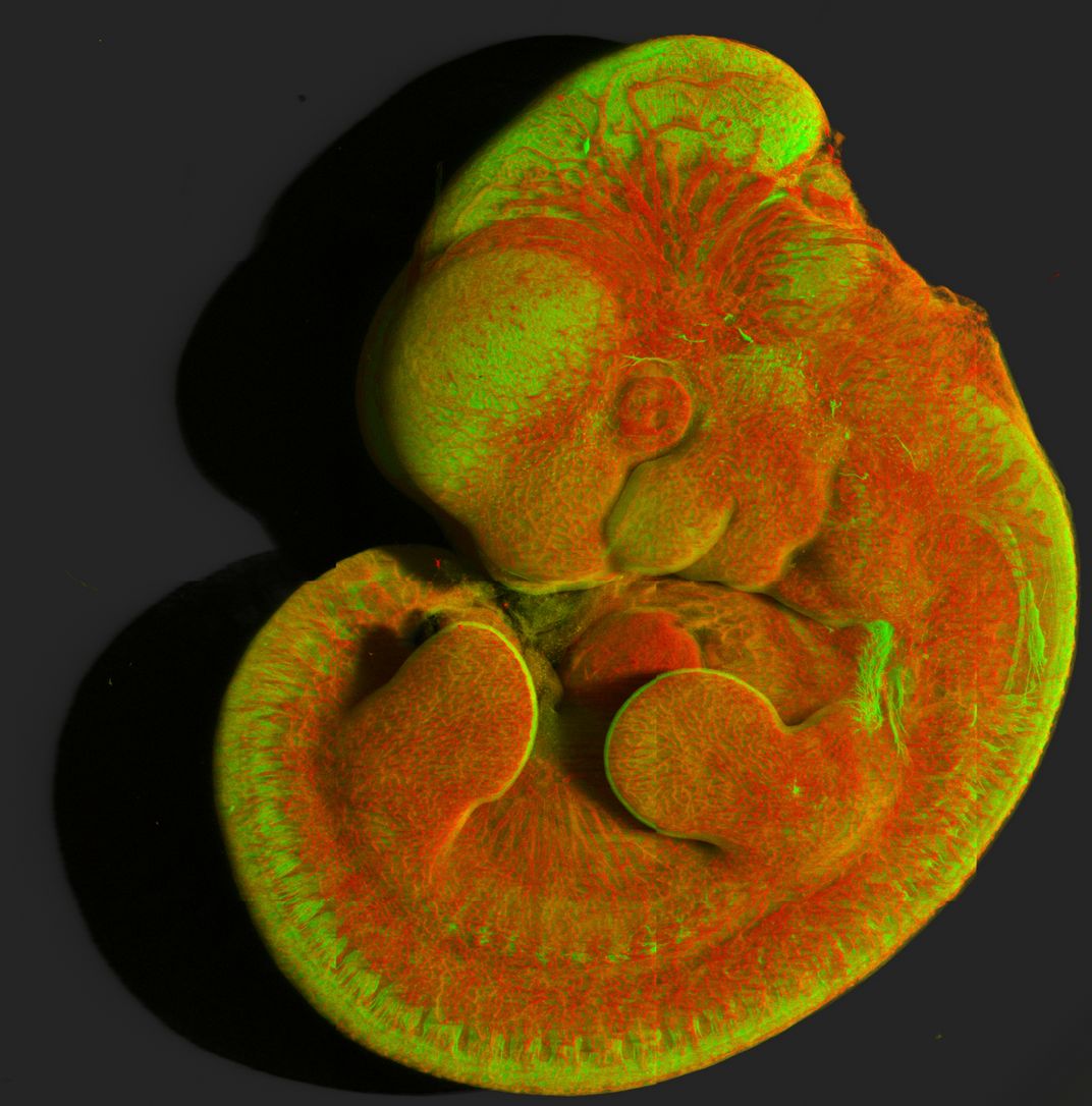
Mouse embryo vasculature (red) and nervous system (green)
Image represents a 3D reconstruction of whole E11.5 mouse embryo vasculature (red) fluorescence visualized via CD31 (a marker for vascular endothelial cells) and nervous system (green) visualized by Tuj1 (Neuron-specific Class III β-tubulin, a marker for neuronal cell bodies and axons), cleared using iDISCO, and captured using Zeiss 880 Confocal Microscope (10X, 0.5NA) stitching tiled confocal images over whole 1.3mm depth. Images were processed for visualization using Imaris software.
Photo Detail
| Date Taken: | 12.2019 |
| Date Uploaded: | 11.2021 |
| Photo Location: | Bethesda, Maryland, United States of America |
| Copyright: | © Daniela Malide |